Medisage Medical Medisage Medical

Top Ways to Ensure Stability in Bone Reconstruction?

Time:2026-09-16 Author:Oliver
0%

Bone reconstruction is not stable simply because a graft fills a defect. Stability depends on fixation, biology, alignment, loading, and patient-specific healing capacity. The Global Burden of Disease Study 2019 reported about 178 million new fractures worldwide in one year. Its findings show the scale of injuries requiring reliable reconstruction. However, fracture numbers alone cannot explain clinical success.

So, how to ensure stability in bone reconstruction? A practical answer begins with careful imaging, stable fixation, controlled micromotion, and preservation of local blood supply. Surgeons must assess bone quality, defect geometry, soft-tissue coverage, infection risk, and the patient’s ability to follow rehabilitation instructions. The AO Foundation repeatedly emphasizes mechanical stability and biological viability as connected treatment priorities. One cannot safely replace the other.

Biology still matters most.

As Gavriil Ilizarov stated, “The blood supply is the most important factor in bone healing.” This principle remains relevant when selecting grafts, planning osteotomies, and protecting periosteal tissues. Recent orthopedic literature also supports individualized fixation and staged reconstruction for complex defects. Yet, clinical decisions are rarely perfect. A rigid construct may protect alignment but restrict useful load transfer. A biologically attractive graft may fail under excessive motion. That tension deserves honest review.

This guide examines fixation strategy, graft selection, imaging, load management, infection prevention, and follow-up evidence. It draws on AO Foundation guidance, peer-reviewed orthopedic research, and the Global Burden of Disease Study. The goal is not a universal formula. It is a safer framework for making stability visible, measurable, and revisable.
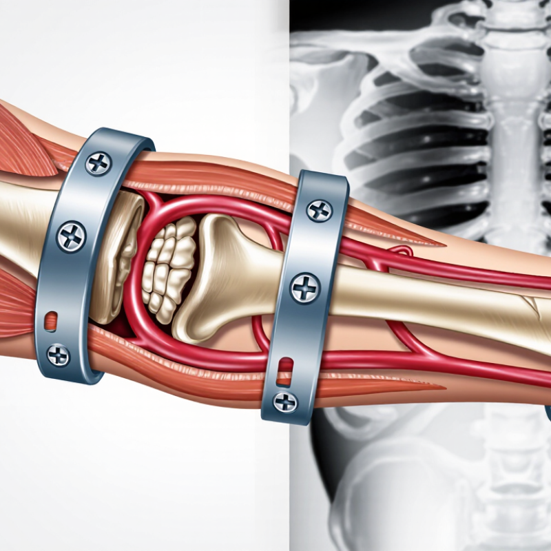
Top Ways to Ensure Stability in Bone Reconstruction?

Understanding the Principles of Stability in Bone Reconstruction

Understanding stability in bone reconstruction begins with a simple distinction: mechanical support and biological healing must work together. A rigid construct can still fail when blood supply is damaged or bone contact is poor. Conversely, controlled movement may encourage healing in selected situations. The correct balance depends on fracture pattern, bone quality, soft-tissue condition, and the patient’s health. There is no universal setting.

Surgeons assess alignment in three dimensions, then choose fixation that matches the expected load. Stable contact reduces unwanted motion at the reconstruction site. Preserving periosteal blood flow protects the cells needed for repair. Careful handling matters. In practice, stability also depends on screw placement, graft quality, and closure of healthy soft tissue. Imaging during surgery can reveal gaps that direct inspection misses. Yet an apparently perfect image does not guarantee biological success.

After surgery, gradual loading should follow healing evidence and clinical assessment, not impatience. Follow-up imaging, pain trends, swelling, and function help identify delayed healing or loss of alignment. Patient factors matter too, including nutrition, smoking, diabetes control, and adherence to movement restrictions. No plan is flawless. Unexpected loading, infection, or weak bone can change the risk quickly. Teams should reassess rather than defend the original decision. This reflective approach makes stability a continuing process, not a single moment in the operating room.

Assessing Bone Quality, Defects, and Reconstruction Requirements

Top Ways to Ensure Stability in Bone Reconstruction?

Stability begins before choosing a graft or fixation method. The key question is whether the remaining bone can carry load. Assess cortical thickness, trabecular structure, density, vascularity, and infection risk. Combine patient history, physical examination, radiographs, and three-dimensional imaging.

The International Osteoporosis Foundation reports that one in three women and one in five men over 50 may experience an osteoporotic fracture during their lifetime. Bone quality is not a minor detail.

Defect assessment should describe more than length and width. Record contained or segmental loss, wall integrity, soft-tissue coverage, alignment, and the distance between viable bone ends.

The Global Burden of Disease 2019 study estimated 178 million new fractures worldwide in 2019. This scale shows the importance of consistent evaluation.

Yet population data cannot predict one patient’s healing response. That is where judgment matters.

Reconstruction must match biology with mechanics.

A contained defect may require a different approach from critical-size segmental loss. The plan should restore length, rotation, joint congruity, and controlled load transfer.

Consider bone density, patient activity, nutrition, smoking status, and expected compliance. Document the reasoning. Reassess early.

Some plans look sound on imaging but fail under real loading conditions. That uncomfortable possibility deserves attention.

Selecting Materials and Fixation Methods for Structural Support

Stable bone reconstruction begins with matching the material to the defect, not choosing the strongest option automatically. Autograft remains valuable because it provides living cells and native growth factors. However, donor-site pain and limited volume can restrict its use. Structural allograft offers shape and support, but incorporation may be slower and less predictable.

A 2023 systematic review in Injury reported nonunion rates of roughly 5–10% across many long-bone fracture cohorts. Critical-size defects showed higher risk. These figures make fixation strategy important. Locking plates can preserve alignment around fragile bone, while intramedullary nails support load sharing along the mechanical axis. External fixation remains useful when swelling, contamination, or soft-tissue loss complicates internal fixation. The AO Surgery Reference advises that fixation must respect biology, not only mechanical stiffness. Excessive rigidity can limit beneficial load transfer.

Surgeons should assess defect length, bone quality, alignment, and expected loading before selecting a construct. The UK National Joint Registry’s 2024 report tracks more than three million procedures, illustrating the value of long-term outcome surveillance. Reconstruction data need the same discipline. Early radiographs can look excellent while delayed union develops later. I have seen planning assumptions fail when patient activity, smoking exposure, or soft-tissue healing changed. That uncertainty deserves explicit documentation. Material choice should be reviewed alongside fixation stability, biological readiness, and the patient’s actual rehabilitation demands.

Top Ways to Ensure Stability in Bone Reconstruction? - Selecting Materials and Fixation Methods for Structural Support

Material or Fixation Method Primary Structural Role Key Advantages Important Limitations Common Clinical Use Stability Considerations
Autologous corticocancellous bone graft Biological repair with limited structural support Contains the patient's own bone cells and matrix; provides osteogenic, osteoinductive, and osteoconductive potential Limited quantity; donor-site pain, infection, bleeding, or sensory complications may occur; not suitable as the sole support for major load-bearing defects Small to moderate defects, nonunion augmentation, and biologic enhancement around fixation Requires stable fixation and adequate vascularized contact with the host bone
Structural cortical allograft Bulk structural reconstruction and load transfer Provides a shaped cortical framework; useful when the defect is too large for autograft Incorporation is slower and variable; risk of fracture, resorption, nonunion, and infection remains; biological remodeling is limited compared with autograft Large segmental defects, revision reconstruction, and selected tumor or trauma cases Needs intimate host-bone contact, secure fixation, and protection from excessive early loading
Demineralized bone matrix Biological stimulation and defect filling Moldable and easier to place in irregular spaces; may provide osteoconductive matrix and variable osteoinductive activity Usually lacks meaningful load-bearing strength; activity varies by preparation and processing Adjunct to fixation, nonunion treatment, and contained defects Should not substitute for a mechanical construct when the defect is exposed to substantial load
Calcium phosphate or beta-tricalcium phosphate ceramic Osteoconductive void filling with gradual resorption, depending on composition Mineral composition resembles bone; available as granules, blocks, or injectable formulations; does not require a donor site Brittle and weak in tension and bending; handling and resorption rates vary; generally unsuitable as an unsupported load-bearing implant Contained cancellous defects and augmentation around a mechanically stable construct Requires containment and protection from shear, bending, and excessive compression
Titanium alloy fixation Primary mechanical stabilization through plates, screws, cages, or rods High strength-to-weight ratio, corrosion resistance, and generally good biocompatibility; produces less imaging artifact than many stainless-steel constructs Stress shielding may occur; fixation can loosen or fatigue-fail; MRI safety depends on the complete implant system Internal fixation for long-bone, pelvic, spinal, and maxillofacial reconstruction Stability depends on screw purchase, plate or rod contour, construct length, fracture gap, and expected loading
Stainless-steel fixation Rigid internal fixation and temporary or permanent load sharing High strength, established surgical handling characteristics, and broad use in plates, screws, wires, and external-fixation components Higher elastic modulus than bone may contribute to stress shielding; imaging artifact and corrosion considerations depend on alloy and environment Trauma fixation, cerclage, wires, and selected reconstructive procedures Avoid mixing incompatible metals when galvanic corrosion is a concern; verify implant-specific MRI conditions
PEEK structural implant Radiolucent structural spacer or cage with controlled load transfer Elastic modulus closer to cortical bone than metals; limited imaging artifact; useful for assessing fusion or bone continuity Biologically inert unless modified or combined with graft; subsidence, migration, and inadequate integration can occur Selected spinal, cranial, or other reconstructive applications requiring radiographic visibility of surrounding bone Requires appropriate endplate or host-bone contact, supplemental fixation when indicated, and control of implant migration
Bone cement, such as PMMA Immediate mechanical interlock or defect filling rather than biologic bone replacement Provides rapid fixation and can fill irregular spaces; does not depend on bone ingrowth for initial stability Nonresorbable; lacks biologic integration; polymerization generates heat; loosening, wear, and cement-related complications are possible Selected joint reconstruction, revision procedures, and specific stabilization or augmentation techniques Adequate cement-bone interdigitation and correct preparation are essential; cement should not replace fixation when structural instability persists
Compression plate with bicortical screws Direct fracture compression and control of bending, rotation, and shear Can provide high initial stability when bone contact and screw purchase are adequate Requires sufficient bone quality and access; excessive periosteal disruption or overly rigid fixation may affect biology Simple transverse or short-oblique fractures and selected reconstructive osteotomies Compression should be applied without creating malalignment; screw distribution and plate working length influence fatigue resistance
Locking plate construct Fixed-angle support and bridge fixation across comminuted or weak bone Maintains angular stability; useful in osteoporotic bone and metaphyseal or periarticular reconstruction Can be too stiff if overbuilt; screw failure, plate fatigue, and nonunion may occur; does not automatically provide compression Comminuted fractures, periarticular defects, and compromised bone stock Balance construct stiffness with controlled micromotion; use suitable plate length and avoid excessive screw density when bridging is intended
Intramedullary nail Load-sharing stabilization along the mechanical axis of a long bone Efficient control of bending; generally preserves soft-tissue coverage and permits early mobilization in appropriate cases May provide limited control of rotation or length in highly comminuted or metaphyseal defects; insertion-related complications are possible Diaphyseal fractures and selected segmental or reconstructive long-bone procedures Correct nail diameter, length, locking pattern, alignment, and fracture reduction are critical to prevent migration or fatigue failure
External fixation Adjustable stabilization outside the injured zone Useful when soft tissues are compromised; permits wound access and can be adapted for gradual deformity correction or bone transport Pin-site infection, frame discomfort, joint stiffness, and loss of rigidity may occur; requires careful follow-up and maintenance Open injuries, infection, severe swelling, limb-length discrepancy, and complex segmental defects Pin spread, frame geometry, bar stiffness, and periodic tightening or adjustment affect construct stability
Clinical selection principle: Material choice should match the defect's size, location, load environment, bone quality, soft-tissue condition, infection risk, and biological healing potential. Structural grafts and fixation devices provide different functions and are often combined rather than used as substitutes for one another.

Applying Surgical Techniques to Maintain Alignment and Stability

Stable bone reconstruction begins with accurate planning, not hardware selection alone. CT images help define defects, fracture lines, and the intended mechanical axis. In the operating room, temporary reduction tools can restore length and rotation before definitive fixation. Alignment should be checked in more than one plane. A straight view can hide a twist. This is easy to miss.

Surgeons commonly use guidewires, reduction clamps, and staged fixation to control the construct. Compression across a suitable fracture line may improve contact, while bridging techniques can protect fragile bone. Screw placement should respect the planned axis and preserve viable soft tissue. Plates, nails, or external supports must match the patient’s anatomy and loading needs. Stability is not the same as rigidity. Excessive force can damage bone or circulation.

Intraoperative imaging provides an important reality check. Comparing limb length, joint orientation, and rotation with the uninjured side can expose subtle errors. Postoperative radiographs then monitor alignment during healing. Weight-bearing decisions should follow fixation strength, bone quality, and healing progress, not impatience. Even experienced teams can overlook a millimeter or a misplaced angle. Reviewing these misses honestly improves future planning. Patient-specific risks, including smoking, infection, and poor nutrition, also deserve active management through coordinated follow-up.

Top Ways to Ensure Stability in Bone Reconstruction

Applying surgical techniques to maintain alignment and stability requires controlling deformity in multiple planes. The chart shows commonly used maximum alignment tolerances for adult tibial shaft fracture fixation.

Clinical interpretation: Coronal alignment is commonly kept within 5°, while sagittal and rotational deformity are commonly limited to 10°. Surgeons support these targets through accurate reduction, provisional fixation, appropriate plate or intramedullary nail positioning, balanced screw placement, fluoroscopic assessment, and protection of the biological environment. Shortening is also commonly limited to approximately 1 cm, although acceptable limits vary by bone, fracture pattern, patient age, and clinical context.

Monitoring Healing and Managing Factors That Affect Reconstruction Stability

Top Ways to Ensure Stability in Bone Reconstruction?

Bone reconstruction stability is built over time, not confirmed on surgery day. Regular reviews help clinicians track pain, swelling, bite, movement, and wound changes. Small shifts can matter. Imaging should match the clinical picture, rather than replace it. Early X-rays may look reassuring while biological healing remains incomplete. A stable reconstruction is not always a quiet one.

Follow-up schedules should reflect the reconstruction site, graft type, general health, and surgical complexity. Clinicians may compare imaging across visits to assess bridging bone, alignment, and hardware position. They also check whether function is improving without excessive load. A patient’s report matters. Persistent tenderness during chewing or a new clicking sound deserves attention, even when images appear acceptable. Healing is individual.

Stability can be influenced by smoking, poorly controlled diabetes, inadequate protein intake, infection, and premature pressure on the area. Medication history and previous healing problems should be discussed openly. Gentle hygiene, prescribed activity limits, and adequate nutrition support repair, but they cannot guarantee success. Follow-up plans sometimes need revision. That is not failure. It is a reason to reassess the biology and mechanics before damage progresses. Missed appointments can hide important changes. Patients should ask which symptoms require urgent contact and when normal activity can resume.

FAQS

What creates stability during bone reconstruction?

Mechanical support and biological healing must cooperate. Good alignment, bone contact, blood flow, and controlled loading all matter. A stiff construct alone cannot guarantee healing.

Why is preserving blood supply important?

Healing cells need adequate blood flow. Rough tissue handling can damage the periosteum and nearby soft tissue. This may weaken biological repair, even when fixation looks secure.

How do surgeons choose a fixation method?

They consider fracture shape, defect length, bone quality, and expected loading. Fragile bone may benefit from locking plates. Load-sharing devices may suit aligned long-bone defects. External support can help when swelling or soft-tissue damage is present.

Is the strongest fixation always the best choice?

No. Excessive rigidity may reduce useful load transfer. Some controlled movement can support healing in selected cases. The correct balance remains patient-specific.

What are the differences between common graft materials?

An autograft contains living cells and natural growth factors. However, it can cause donor-site pain and provide limited volume. A structural allograft offers shape and support. Its incorporation may be slower and less predictable.

Can a good surgical image prove successful healing?

No. Imaging may show excellent alignment while healing remains delayed. Blood supply, bone quality, soft-tissue recovery, and patient activity still matter. A small unseen gap can change the outcome.

What should be monitored after reconstruction?

Follow-up images, pain, swelling, movement, and daily function provide useful clues. A sudden increase in pain or swelling deserves clinical review. Loss of alignment may appear gradually. Early reassurance can sometimes be wrong.

How do patient habits affect reconstruction stability?

Nutrition, smoking exposure, diabetes control, and activity restrictions influence healing. Unexpected loading can stress the reconstruction before the bone is ready. Recovery plans must match real behavior, not ideal behavior. That assumption sometimes fails.

Why should the treatment plan be reassessed?

Infection, weak bone, delayed healing, or changing activity can alter risk quickly. Teams should question the original plan when new evidence appears. No plan is flawless. Stability is a continuing process, not one operating-room moment.

Conclusion

Bone reconstruction depends on achieving reliable stability from the earliest stage of treatment. The key to understanding how to ensure stability in bone reconstruction is to evaluate bone quality, defect size, location, and the mechanical demands placed on the affected area. These factors guide the choice of structural support materials and fixation methods, helping restore alignment while providing enough strength for healing. A carefully planned reconstruction should balance rigidity with appropriate biological conditions so that new bone can develop effectively.

During surgery, accurate positioning, secure fixation, and protection of surrounding tissues are essential for maintaining stability. Afterward, healing should be monitored through clinical assessments and imaging, with attention to movement, loading, nutrition, infection prevention, and other factors that may influence recovery. Early recognition of instability or delayed healing allows timely adjustment of treatment and supports a safer, more predictable reconstruction process.

Oliver

Oliver

Oliver is a seasoned marketing professional with a wealth of expertise in driving brand awareness and engagement. With a deep understanding of our company's product offerings, he consistently delivers high-quality content that enriches our professional blog. His insights not only shed light on......